Research Interests
Structural and Functional Interrogation of Bacterial Antiviral Defense Systems: evolution of novel defense strategies in prokaryotes
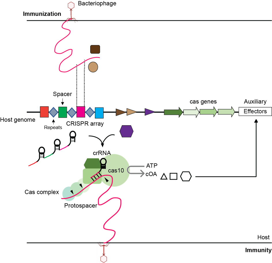
My research is centered upon the mechanistic understanding of the diverse spectra of bacterial defense systems at the molecular
level. On an elaborate note, I ‘select’ single or multi-component novel defense systems in prokaryotes, 'explore' their biophysical properties,
'map' their interactions with small signal molecules, nucleic acids or other partner proteins, 'see' the relevant states of these systems at atomic-resolution
using single particle cryo-EM and X-ray crystallography and finally utilize structure guided biochemical functional analysis to 'dissect' their mechanism of
action.
To my belief, this combinatorial approach is quite powerful for solving complex
biological processes. My research area involves strong understanding of structural biology, biochemistry and biophysical properties of membrane proteins,
multicomponent systems, and filament proteins. I utilize sophisticated tools like cryo-electron microscope and x-ray crystallography to achieve atomic-resolution
structures of the proteins and follow different approach like generating nanobodies or antibody fragments to aid the structural studies on the target of interest.
I design and perform exciting biochemical experiments to establish structure-function correlation of the complex biological systems.
Currently my work is focused on CRISPR associated effector proteins involved in novel defense strategies
like membrane-depolarization, depletion of essential cellular molecule and hydrolysis of essential component of the cell by filament formation. My doctoral dissertation was
focused on membrane spanning antibacterial efflux transporter QacA, whose structure was determined with bound nanobodies, and mechanism of action was deciphered by structure
guided biochemical studies. Please look into the recent research highlights below and Publication to know more.
Recent Research Results
We seek to understand the diversity and versatility of defense strategies underlying the arms race between viruses and bacterial surveillance systems by focusing on cyclic-oligoadenylate (cOA) activated auxiliary CARF-effector proteins associated with the type-III CRISPR-Cas system. The predominantly characterized CARF-associated cOA-activated effector domains act as nucleases to cleave DNA and/or RNA in a non-specific manner. However, the CARF domain containing auxiliary effector proteins associated with transmembrane domains or different enzymatic domains other than nucleases are poorly understood.
TYPE III CRISPR ASSOCIATED CARF-TIR 1 (Cat1)
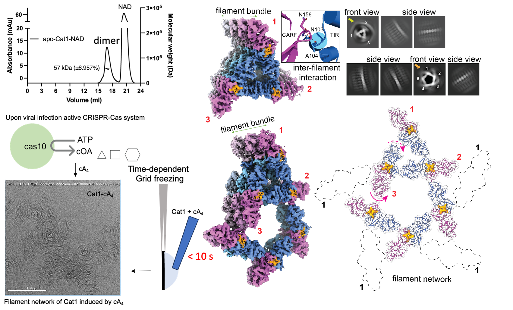
We extended our studies on CRISPR associated CARF-TIR (Toll/Interleukin-1 Receptor) effectors. TIR domains are found in both eukaryotes and
prokaryotes and are involved in innate immune systems and cell death responses via homotypic interactions or self-association with other TIR-domains. It is of mechanistic
interest to elucidate whether and how TIR domain containing CARF effectors are enhancing the immunity of the bacterial adaptive CRISPR defense systems. The biochemical
analysis and cryo-EM studies reveal that even though the apo CARF-TIR (Cat1) protein elutes as a dimer, the effector forms a triple filament bundle which further
associates to form a supramolecular pentameric filament bundle in presence of cA4 signal molecule during CRISPR defense. The cryo-EM structures of the cA4 bound Cat1
unravel a unique filament assembly where the dimeric CARF-domains form the center of the filament, and the TIR-domains are radially arranged. The cA4 binding pocket
is formed by two such Cat1 dimers associated in head to tail fashion where the bottom CARF-dimer forms the base of the pocket and the top CARF dimer caps the bound cA4.
Hence, the two Cat1 dimers forms the repeat units of the filament assembly. Moreover, the cryo-EM structures of cA4 bound Cat1 with its substrate analogue and the
product reveals that the NADase catalytic site of the TIR domain is formed by two TIR domains arranged in head to tail manner as a part of two Cat1 dimers and the
finding was further verified by systematic mutation based functional studies by the Marraffini group. All together our study provides a mechanistic basis of Cat1
filament assembly in presence of the signal molecule and elucidates NAD depletion as a novel defense strategy by CRISPR systems against phages. This work on a series
of type-III CRISPR-associated CARF-effector proteins can establish a framework to understand the evolution of bacterial adaptive immune strategies. To know more,
please see our 2025 paper in Science.
TYPE III CRISPR ASSOCIATED CARF-Adenosine Deaminase 1 (Cad1)
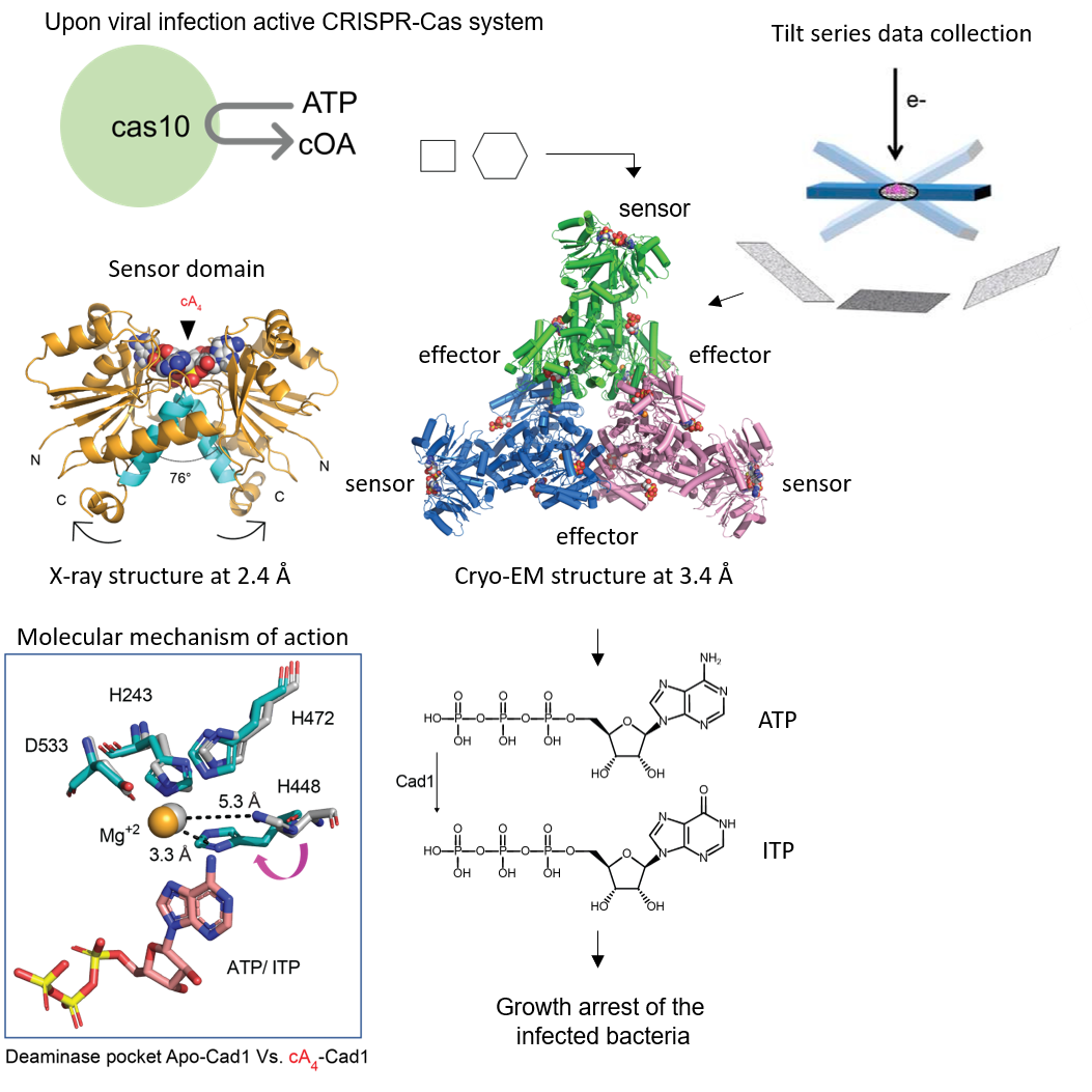
Cad1 (CRISPR-associated adenosine deaminase 1) contains deaminase enzyme domain which is repurposed to antiphage defense. I employed
biophysical characterization and biochemical assays to find the signal cOAn molecules that activate Cad1 and to reveal its oligomeric states. I performed
x-ray crystallography to solve the structure of the CARF domain of Cad1 in the apo, cA4 and cA6 bound forms at 3.6 Å, 2.4 Å and 1.8 Å resolution respectively
revealing the mechanism of signal recognition by the dimeric CARF domains during CRISPR antiphage defense. The cryo-EM structure of the full-length Cad1 in the apo state,
the ATP-bound state and cA 4 bound state in presence of ATP at 3.4 Å, 3.6 Å and 3.2 Å resolution respectively along with collaborative in vivo functional studies in the
Marraffini lab revealed that Cad1 is a hexameric, cA4/cA6 activated, metal dependent, ATP to ITP deaminase. Additionally, the ATP bound apo and cA4 -bound Cad1 cryo-EM structures lead to the identification of a unique ATP binding site at the interface of the CARF and deaminase domains which is required for activity.
This pocket binds double the number of ATPs in the cA4 -bound structure indicating its involvement in ATP sequestration upon signal recognition. Cad1 mediated ATP depletion and
ITP accumulation leads to growth arrest of the phage infected bacteria and stops the spread of viral particles to the rest of the population. To know more, please see our 2024 paper
in Cell.
TYPE III CRISPR ASSOCIATED membrane protein 1 (Cam1)
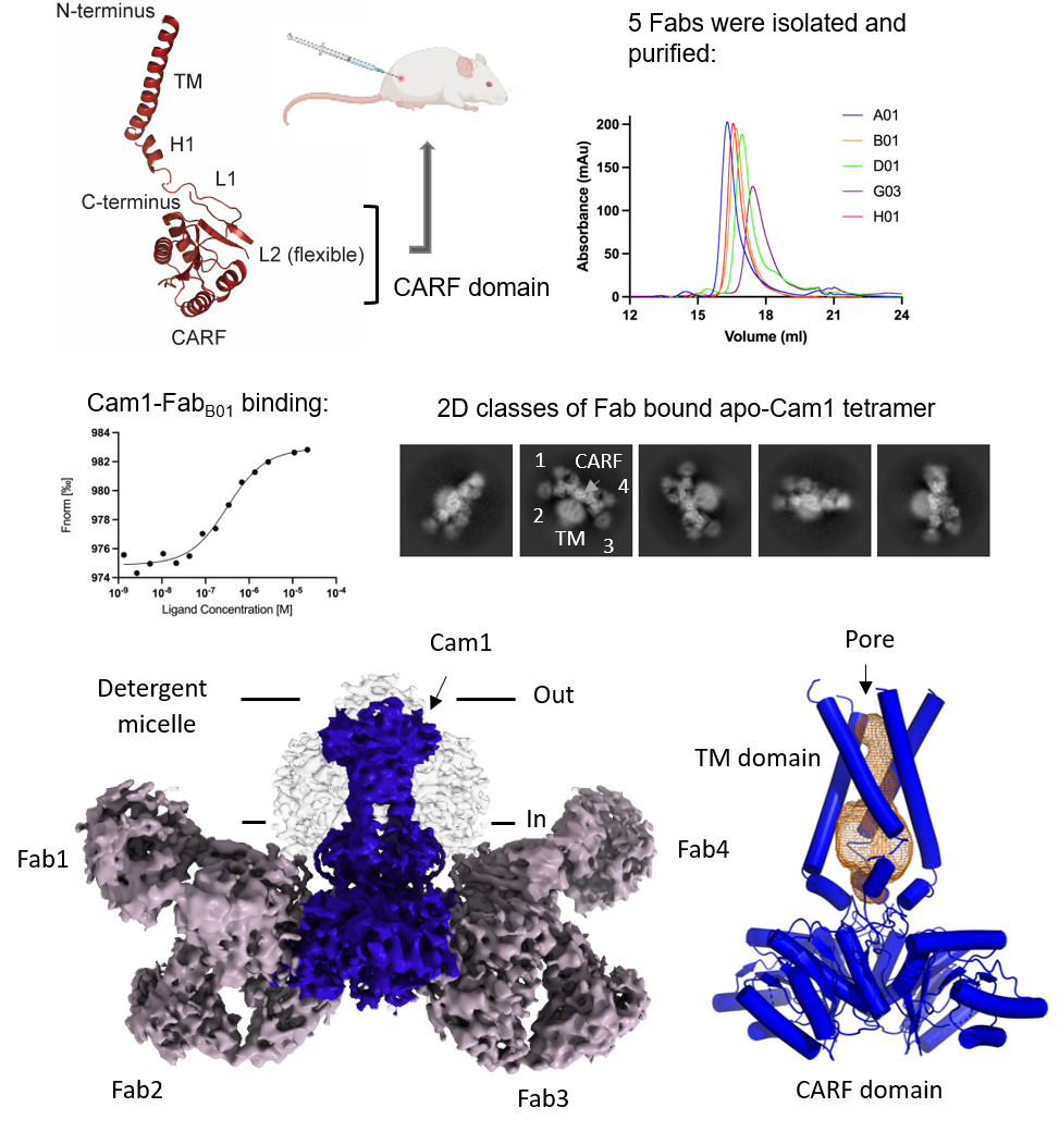
Type-III CRISPR-Cas associated Cam1 effector contains an N-terminal transmembrane helix linked to the C-terminal CARF domain.
Our recent work identified that Cam1 oligomerizes to a homotetramer and causes abortive infection by membrane-depolarization following cA4 binding to its
CARF domain during CRISPR-Cas immunity. To elucidate the molecular basis of membrane-depolarization by Cam1, we employed state-of the art single particle
cryo-electron microscopy (cryo-EM) to solve the atomic resolution structure of full-length Cam1. A key barrier to progress has been the inherent toxicity
involved with Cam1 heterologous expression in bacterial systems and low molecular weight of the protein (molecular weight of Cam1 monomer is 25 kDa) for
single particle cryo-EM studies. The challenge with the low yield of the protein was overcome by increasing the volume of bacterial expression system and
employing suitable detergent and lipids with high extractability to solubilize the protein from the bacterial membrane. To address the second challenge of
low molecular weight of Cam1, we generated monoclonal antibodies against the CARF domain of Cam1 protein. We used the high affinity Fab fragments of the
monoclonal antibodies as fiducial markers to solve the apo-Cam1 structure by cryo-EM at 3.9 Å resolution. The structure reveals that Cam1 tetramerizes to
form a pore in the membrane and the electrostatics of the residues lining the pore is suggestive of membrane-depolarization by cation transport. To
explore the mechanistic details of activation of Cam1 protein during CRISPR defense, we will employ similar approach to solve the structure of Cam1
protein in the presence of the activator molecule, cA4. The proposed study should highlight the fundamental molecular mechanism of cOA-activated membrane
associated effectors that belongs to the CRISPR-Cas systems. To know more, please see our 2024 paper in
Nature.
Antibacterial efflux transporter QacA (Quaternary Ammonium Compound Transporter A)
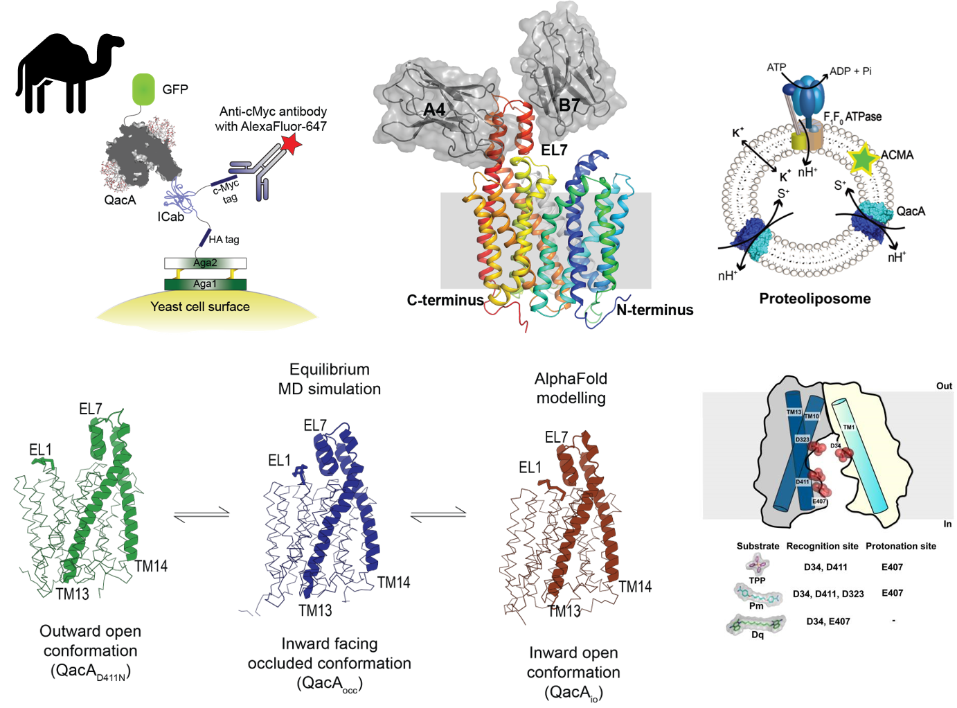
Efflux of antibacterial compounds is a major mechanism for developing antimicrobial resistance. In the Gram‐positive pathogen
Staphylococcus aureus, QacA, a 14 transmembrane helix containing major facilitator superfamily antiporter, mediates proton‐coupled efflux of mono and divalent cationic
antibacterial compounds. In this study, we report the cryo‐EM structure of QacA, with a single mutation D411N that improves homogeneity and retains efflux activity against
divalent cationic compounds like dequalinium and chlorhexidine. The structure of substrate‐free QacA, complexed to two single‐domain camelid antibodies, was elucidated to
a resolution of 3.6 Å. The structure displays an outward‐open conformation with an extracellular helical hairpin loop (EL7) between transmembrane helices 13 and 14,
which is conserved in a subset of DHA2 transporters. Removal of the EL7 hairpin loop or disrupting the interface formed between EL7 and EL1 compromises efflux activity.
Chimeric constructs of QacA with a helical hairpin and EL1 grafted from other DHA2 members, LfrA and SmvA, restore activity in the EL7 deleted QacA revealing the allosteric
and vital role of EL7 hairpin in antibacterial efflux in QacA and related members.
Systematic mutagenesis resulted in the identification of D34 (TM1), and a cluster of acidic residues in TM13 including E407 and D411 and D323 in TM10, as being crucial for
substrate recognition and transport of monovalent and divalent cationic antibacterial compounds. The transport and binding properties of QacA and its mutants were explored
using whole cells, inside-out vesicles, substrate-induced H+ release and microscale thermophoresis-based assays. The activity of purified QacA was also observed using
proteoliposome-based substrate-induced H+ transport assay. Our results identify two sites, D34 and D411 as vital players in substrate recognition, while E407 facilitates
substrate efflux as a protonation site. We also observe that E407 plays an additional role as a substrate recognition site for the transport of dequalinium, a divalent
quaternary ammonium compound. These observations rationalize the promiscuity of QacA for diverse substrates. The study unravels the role of acidic residues in QacA with
implications for substrate recognition, promiscuity and processive transport in multidrug efflux transporters, related to QacA. To know more, please see our 2023 paper in
The EMBO Journal and 2019 paper in
Journal of Molecular Biology.